Joseph Goldstein
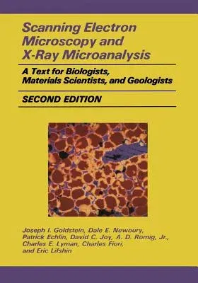
(Author)Scanning Electron Microscopy and X-Ray Microanalysis: A Text for Biologists, Materials Scientists, and Geologists (1992. Softcover Reprint of the OrigPaperback - 1992. Softcover Reprint of the Original 2nd 1992, 28 September 2011

Qty
1
Turbo
Ships in 2 - 3 days
In Stock
Free Delivery
Cash on Delivery
15 Days
Free Returns
Secure Checkout
Print Length
840 pages
Language
English
Publisher
Springer
Date Published
28 Sep 2011
ISBN-10
1461276535
ISBN-13
9781461276531
Description
Product Details
Authors:
Book Edition:
1992. Softcover Reprint of the Original 2nd 1992
Book Format:
Paperback
Country of Origin:
NL
Date Published:
28 September 2011
Dimensions:
25.4 x
17.78 x
4.24 cm
ISBN-10:
1461276535
ISBN-13:
9781461276531
Language:
English
Location:
New York, NY
Pages:
840
Publisher:
Weight:
1433.35 gm

