Farhood Saremi
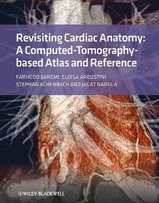
(Author)Revisiting Cardiac Anatomy: A Computed-Tomography-Based Atlas and ReferenceHardcover, 15 November 2010

Temporarily out of stock
Free Delivery
Cash on Delivery
15 Days
Free Returns
Secure Checkout

Print Length
320 pages
Language
English
Publisher
Wiley-Blackwell
Date Published
15 Nov 2010
ISBN-10
1405194693
ISBN-13
9781405194693
Description
Product Details
Author:
Book Format:
Hardcover
Country of Origin:
GB
Date Published:
15 November 2010
Dimensions:
27.94 x
21.59 x
2.29 cm
ISBN-10:
1405194693
ISBN-13:
9781405194693
Language:
English
Location:
Chichester, England
Pages:
320
Publisher:
Weight:
1224.7 gm