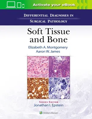
New in the Differential Diagnosis in Surgical Pathology series, this
abundantly illustrated title helps you systematically solve tough
diagnostic challenges in soft tissue and bone pathology. It uses select
images of clinical and pathological findings, together with succinct,
expert instructions and diagnostic pearls, to guide you through the
decision-making process by distinguishing between commonly confused
lesions of soft tissue and bone. By presenting material according to the
way pathologists actually work, this user-friendly volume helps you
quickly differentiate entities that have overlapping morphologic
features.
- Presents over 130 differential diagnoses in soft tissue and bone
pathology, including the most common entities as well as selected rare
diseases.
- Provides concise, bulleted summaries of clinical and pathological
findings and relevant pictorial examples on the corresponding pages.
- Expertly guides you in using hematoxylin and eosin-stained sections as
a tool to better evaluate small samples, precisely guiding ancillary
testing, and using radiologic clues for diagnosis of bone lesions.
- Features over 1,300 high-quality, full-color images of similar-looking
lesions side by side for easy comparison with respect to
clinicopathologic features and ancillary tests.
- Includes sections on soft tissue (Spindle Cell, Adipose Tissue,
Myxoid, Epithelioid, Vasoformative, Pleomorphic, and Round Cell) along
with those in bone (Osteoblastic, Cartilage, Fibroosseous, Fibrous,
Giant cell, Round Cell, Vascular, Cystic, Pleomorphic, Notochordal
Tumors, and Synovial).
- Ideal for practicing pathologists, pathologists in training,
residents, and medical students.
Enrich Your Ebook Reading Experience
- Read directly on your preferred device(s), such as computer,
tablet, or smartphone.
- Easily convert to audiobook, powering your content with natural
language text-to-speech.

