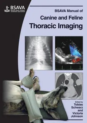
BSAVA Manual of Canine and Feline Thoracic ImagingPaperback, 4 August 2008

Qty
1
Turbo
Ships in 2 - 3 days
Only 2 left
Free Delivery
Cash on Delivery
15 Days
Free Returns
Secure Checkout

Part of Series
BSAVA British Small Animal Veterinary Association
Part of Series
BSAVA Manuals
Print Length
200 pages
Language
English
Publisher
BSAVA
Date Published
4 Aug 2008
ISBN-10
0905214978
ISBN-13
9780905214979
Description
Product Details
Book Format:
Paperback
Country of Origin:
GB
Date Published:
4 August 2008
Dimensions:
29.21 x
20.83 x
2.54 cm
ISBN-10:
0905214978
ISBN-13:
9780905214979
Language:
English
Pages:
200
Publisher:
Weight:
1700.97 gm